Introduction
The disease cataract is the opacification of lenses frequently occurs in diabetic patient with old age because of increased levels of glycosylated haemoglobin are involved in increased risk of cataract formation.1 Many cataract inducing factors have been reported according to the various experimental data; the biochemical reason of cataractogenesis is still not clear till date. The cataract occurs because of multiple factors, mainly happen due to the protein aggregation on the eye lenses. The lenses Na+ — K+ -ATPase activity shows very important action in maintenance of transparent nature of the lens, and its imbalance leads to deposition of Na+ and loss of K+ with water absorption of the lens fibres leading to cataract formation.2 Numbers of drugs have been already used for treatment of cataract but not a single from them proved useful treatment of cataract.3 The aldose reductase is enzyme probably plays a role in the development of this eye problem like cataract.4 The aldose reductase show its action on sugar molecules like glucose, galactose and xylose and convert them into their respective alcohols by biochemical pathways these alcohols are known as polyols accumulate within the lens there by producing osmotic effects. Hence polyols does not have a capacity for diffusing out easily nor metabolizes rapidly and causes hyper tonicity responsible for formation of cataract.5 The oxidative mechanism also plays a crucial role in biological phenomena including cataract formation. The formation of superoxide radicals in the aqueous humour and in lens, lens and its derivatization to other potent oxidants may be responsible for initiating various biochemical toxic reactions leading to formation of cataract.6 Angiotensin converting enzyme inhibitors have been shows protection from free radical damage in many experimental procedures.
Ascorbic acid (ACE Inhibitor) was shown potent anticataract activity in vitro due to antioxidant and free radical scavenging activity.7, 8 Hence we take Ascorbic acid as standard and measure various parameters including (Na + & K+ ) estimation, Na+ -K + ATPase activity, Proteins (total proteins and water soluble proteins) and malondialdehyde (MDA) in vitro on goat lenses.
Materials and Methods
Drugs
Drugs Ascorbic acid, Penicillin and streptomycin were obtained from Loba chemicals, Spectrochem Pvt. Ltd. and some local chemical supplier.
Experimental procedure
Collection of eyeballs
Goat eyeballs used for the study were collected from the local slaughterhouse and stored at 0-40 C.
Lens culture
Fresh goat eyeballs used for the study are withdrawal from slaughterhouse. Artificial aqueous humour is used for anticataract activity. Aqueous humour contains (NaCl) 140 mM, KCl 5mM, MgCl2 2 mM, NaHCO3 0.5 mM, NaHPO4 0.5 mM, CaCl2 0.4 mM and glucose 5.5 mM) at room temperature and maintain pH 7.4 by addition of NaHCO3). Penicillin G and streptomycin 250 mg added for prevention of bacterial growth.
Cataract formation
The glucose solution having concentration 55mM was used for the cataract formation. Higher concentration of glucose metabolizes by the sorbitol pathway. Cataract was formed due to the accumulation of polyol (Sugar + Alcohol) which causes oxidative stress and over-hydration which forms cataract. All these lenses were incubated in aqueous humour which is artificially prepared with different concentration of Glucose for 72 hours.
Study design and groups
Goat lenses were divided into six groups of six lenses each following Table 1
Table 1
Treatment groups for anticataract activity
Homogenate preparation
The homogenate is prepared by 72 hours incubation, homogenate of lenses was prepared by using Tris buffer (0.23 M, pH-7.8) containing 0.25 X10⁻³ sub M EDTA and homogenate makeup up to 10 % w/v. The prepared homogenate was centrifuged at 9,000 G at 4°C for 1 hour and the supernatant for the final solution was isolated from the centrifuge tube which is used for estimation of biochemical parameters. For estimation of water-soluble proteins, homogenate was prepared in sodium phosphate buffer (pH-7.4).
Biochemical analysis
The electrolyte Sodium and Potassium (Na+ and K+) was estimated by using flame photometry. The sodium Potassium ATPase activity was performed by using Unakar and Tsui method9 and estimation of protein was done by Lowry's method.10 The oxidative stress level was analysed by Wilbur’s method.11
Results and Discussion
Photographic evaluation
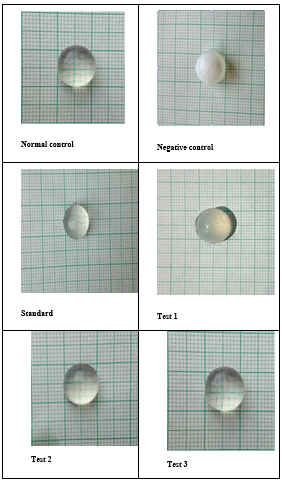
After all experimental procedure lenses were placed on graph paper with the posterior surface touching the graph and observed by using magnifying lens. All the 5.5 mM Glucose (Normal lens), 55 mM Glucose (Cataract induced lens), 40 µg/ml Ascorbic acid (Standard) were analysed by placing on graph paper. All other three lenses treated with Pioglitazone with concentrations of 15, 30 and 60 µg/ml respectively as shown inFigure 1.
Table 2
Effect of pioglitazone on degree of opacity on lens by glucose-Induced cataract
Table 0
Pioglitazone was subjected for in vitro anticataract activity by goat eye lens model. Pioglitazone has multiple biological functions here which play anticataract activity.
Pioglitazone significantly protected the lens morphology and activity and clarity: 50% of the eyes had almost clear lenses; in contrast, 100% of the negative control eyes developed dense nuclear opacity. From the current study, it is evident that Pioglitazone protects the lens against oxidative stress. These results in glucose-induced cataracts in vitro studies not only demonstrate the protective effect of Pioglitazone but also indicate that it prevents cataractogenesis by virtue of its antioxidant properties. Pioglitazone, therefore, may be useful for prophylaxis or therapy against cataract. After 72 hr. of incubation in glucose 55 mM, the lens becomes completely opaque as against lenses in normal control. Incubation of lenses with Pioglitazone and ascorbic acid both the concentrations were used, which seem to retard the progression of opacification compared with lenses incubated in glucose 55 mM (Negative Control). The effect of Pioglitazone on the positive control groups, showed considerable retardation in the progression of lens opacification and which is near normal when compared to negative control.
Table 3
Effect of pioglitazone on protein levels (total proteins and water-soluble proteins) in goat lens homogenate after 72 hours of incubation in glucose 55 mm induced cataract
N=6, values are expressed as Mean ± SEM. Comparison were made as following, # p < 0.05,## p < 0.01 when compared with normal control. * p < 0.05, ** p < 0.01 when compared with negative control. (Values are compared on 72hr by one way ANOVA Dennett test) N. S. — non-significant.
The 55 mM Glucose treated lenses (Group-II) showed significantly low concentrations of proteins (total and water-soluble proteins) in the lens homogenate (P<0.01) compared with normal lenses (Group-I). Ascorbic acid treated lenses (Group-III) and Lenses treated with Pioglitazone (Group-IV, V, VI) showed higher concentrations of proteins (total and water-soluble proteins) (P<0.01) compared with 55 mM Glucose treated lenses (Group-II).
Conclusion
In the present work, Pioglitazone treated group shows the increase in protein content (water-soluble) by prevention of cataractogenesis. This cataract is due to the higher glucose concentration. Pioglitazone shows anticataract activity due to presence of antioxidant activity and prevention of the cataract forming factors.


